Figure 1.

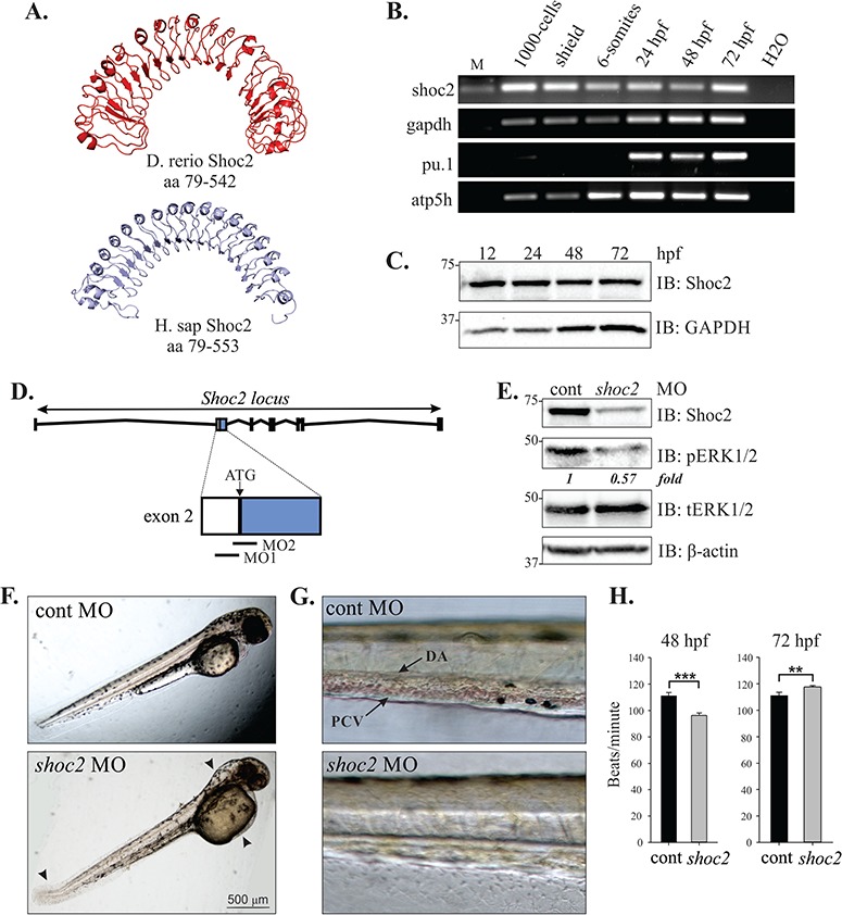
Knockdown of Shoc2 disrupts morphogenesis and causes severe anemia. (A) Homology models of zebrafish and human Shoc2, residues 79-561 and 79-582, respectively, were constructed using the published structure of Arabidopsis Flg22-FLS2-BAK1 immune complex (PDB id: 4MN8) and Leptospira interrogans LRR protein LIC11098 (PDB id: 4U08) as templates for zebrafish and human Shoc2 models correspondingly. The modeling was done using the I-TASSER server and figures were prepared using PyMol software. For modeling detail see (52,53). (B) shoc2 mRNA was detected using reverse transcription polymerase chain reaction (RT-PCR) at the indicated times. pu.1, atp5h and gapdh were used as reference genes. pu.1 was previously shown to express after 16 hpf (54). (C) Western blot analysis of zebrafish embryos. Embryos were harvested for immunoblotting at indicated time points. The expression of Shoc2 and GAPDH was analyzed using specific antibodies. GAPDH was used as a loading control. (D) Schematic representation of Shoc2 loci and MO-targeting sites. (E) Embryos injected with shoc2 and control MO were harvested for immunoblotting at 72 hpf. The expression of indicated proteins was analyzed using specific antibodies. (F) Representative body images of control and shoc2 morphants at 48 hpf (taken from the set of embryos analyzed in E, G and H). One-cell stage embryos were injected with the standard control MO and shoc2 ATG MO1. The optimal dose for microinjection of MO was determined as that which resulted in specific defects but did not cause gross lethality or global defects. As shown, at 48 hpf, 70.4% (57 out of 81) of shoc2 morphants displayed an enlarged hindbrain (arrowhead), enlarged yolk, a posterior kink in the tail (arrow). (G) Representative body images of control and shoc2 morphants at 72 hpf. shoc2 MO-injected embryos showed reduced numbers of cells in circulation when compared to control embryos. (H) The cardiac heart rate of control and shoc2 morphants was measured at 48 and 72 hpf. Significant difference in heart rate was observed in shoc2 morphants when compared to WT. Error bars represent means with SEM. ***P < 0.001 and **P < 0.05 (Student’s t-test). MO: morpholino, hpf: hours post fertilization, M: DNA marker, PVC: posterior cardinal vein, DA: dorsal aorta.
