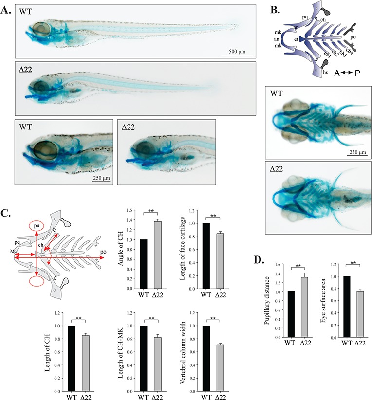
Figure 4.

shoc2 loss leads to defects in craniofacial cartilage specification and differentiation. (A) Lateral and ventral views of a 6 dpf WT and shoc2Δ22 crispant larvae detected by Alcian blue staining. Mutant larvae show significant changes in head cartilage. (B) Schematic representation of the different head cartilage elements in ventral view of 6 dpf. Anterior limit (an), articulation (ar), ceratobranchial pairs 1 to 4 (cb1–4), ceratohyal (ch), ethmoid plate (et), hyosymplectic (hs), Meckel’s cartilage (mk), palatoquadrate (pq), posterior limit (po). (C) Schematic representation of parameters quantified; length of ceratohyal (CH) and CH to Meckel’s cartilage (mk), angle of CH, pupillary distance (pu), eye surface area, width of vertebral column and length of total face cartilage (red arrows) were measured for morphometry. Y-axes on graphs indicate the fold change of shoc2Δ22 crispants compared to WT. (D) Measured distance between the eyes and the eye surface area of 6 dpf WT and shoc2Δ22 crispant larvae. Three biological replicates were performed for all experiments (n = 20/group). (**P < 0.05, Student’s t- test). Error bars represent means with SEM. A: anterior, P: posterior.
