Figure 6.

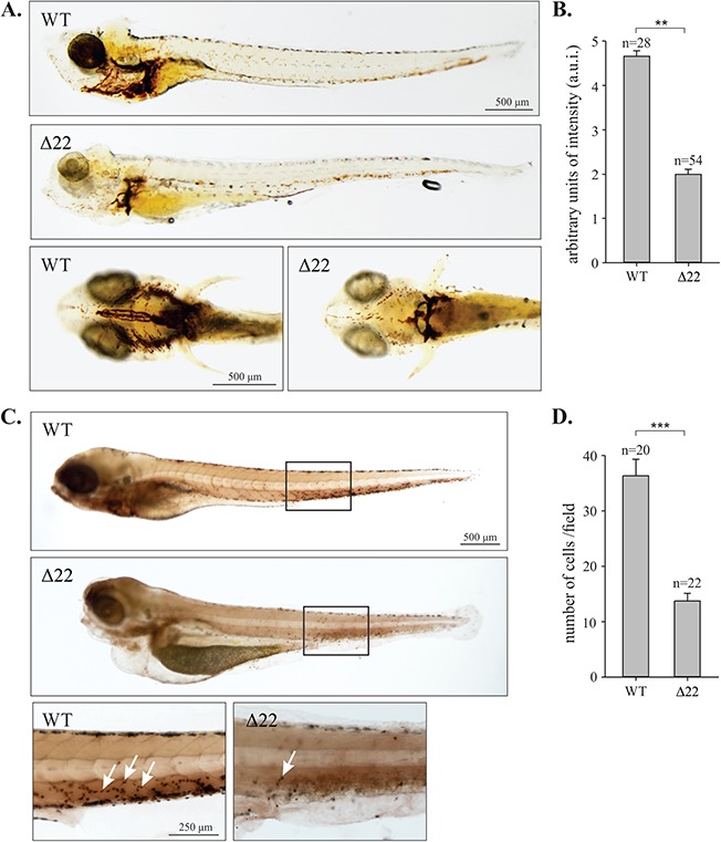
Impaired hematopoiesis in shoc2Δ22-crispant larvae. (A) o-Dianisidine staining detecting hemoglobin of erythropoietic cells in control and shoc2Δ22 larvae at 6 dpf. In control, larvae hemoglobin-positive cells were found on the yolk sac and tail while shoc2Δ22 crispants showed significantly decreased number of cells. Images are shown in lateral and ventral view. (B) Quantitative analysis of the number of erythropoietic cells in circulation. Relative intensity of hemoglobin staining was scored in arbitrary units of intensity (a.u.i.) 0 to 5, 0 being the weakest and 5 the strongest. **P < 0.05 (Student’s t-test). (C) Histochemical staining for mpx enzyme activity in WT and shoc2Δ22 at 6 dpf. Larvae are shown in lateral view with the anterior to the left. The images are representative of ≥ 20 larvae in each group. The staining was repeated with similar results for three independent experiments. Arrows point to mpx-positive cells. (D) The number of mpx-positive cells within the indicated box (five somites). Error bars represent means with SEM. ***P < 0.001 (Student’s t-test).
