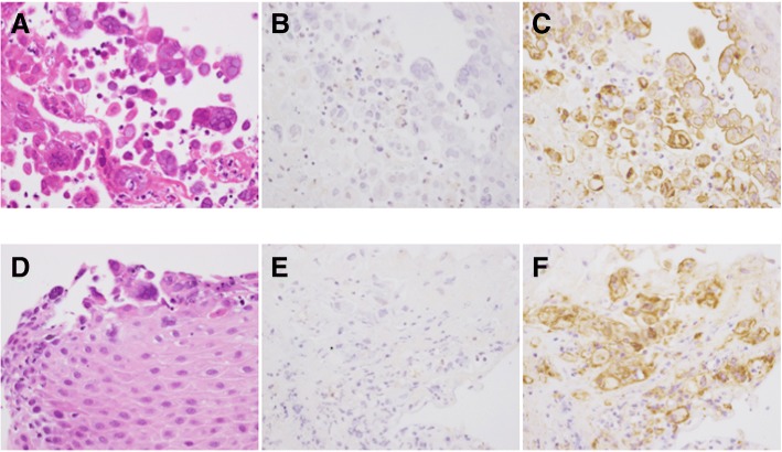
Fig. 3.
Histopathological findings of tonsillar (a-c) and esophageal (d-f) biopsies. a, d: Hematoxylin and eosin staining showing discohesive superficial squamous epithelial cells containing Cowdry type a or type b intranuclear inclusions. b, e: Negative immunohistochemical staining for HSV-1. c, f: Positive immunohistochemical staining for HSV-2

