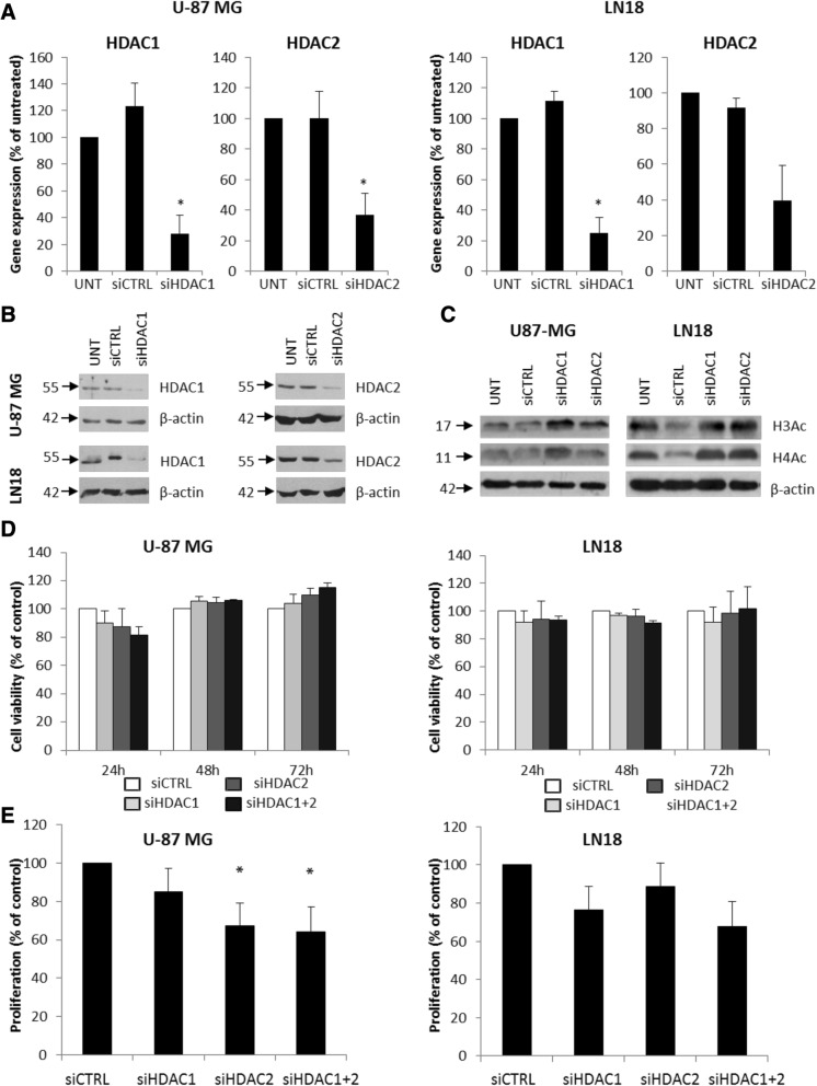
Fig. 3.
Knockdown of HDAC 1 and HDAC 2 results in reduced cell proliferation. a HDAC 1 and HDAC 2 expression was estimated by qRT-PCR in U-87 MG and LN18 cells after gene silencing using specific siRNAs. b Western blot analysis shows efficacy of HDAC 1 and HDAC 2 knockdown at protein level. c Western blot for acetylated histones H3 and H4 (H3Ac, H4Ac) in HDAC 1 and HDAC 2 depleted U-87MG and LN18 cells 48 h after siRNA transfection. d MTT metabolism test for cell viability 24, 48, and 72 h after transfection with HDAC 1 or/and HDAC 2 siRNAs or a control siRNA. e BrdU incorporation test for cell proliferation 48 h after knockdown of HDAC 1 or/and HDAC 2 in U-87MG and LN18 cells. The respective p values were calculated using type 2 two-tailed t test, and p < 0.05 was considered statistically significant. *p value < 0.05, n = 3

