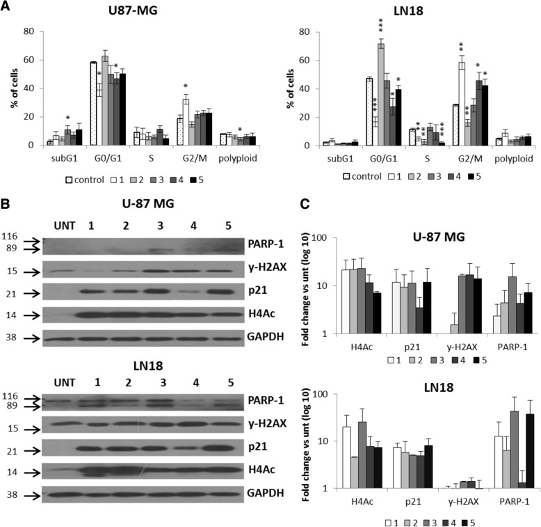
Fig. 5.
Antiproliferative effects of 1–5 on glioma cells. a The effects of compounds 1–5 on cell cycle in glioma cells were determined by propidium iodide (PI) staining and flow cytometry. Quantification of three experiments is presented. The respective p values were calculated using type 2 two-tailed t test, and p < 0.05 was considered statistically significant: *p value < 0.05, **p value < 0.01, ***p value < 0.001. b Total protein extracts were collected from U-87 MG and LN18 cells exposed to HDACi. Representative immunoblot shows results of western blot analysis of PARP-1 cleavage, γ-H2AX, p21, and H4Ac levels in U-87 MG and LN18 cells after exposure to 5 μM 1–3, 5, and 1 μM 4. c Densitometry analysis of western blot of PARP-1 cleavage, γ-H2AX, p21, and H4Ac levels in U-87 MG and LN18 cells after exposure to 5 μM 1–3, 5, and 1 μM 4 (n = 2)

