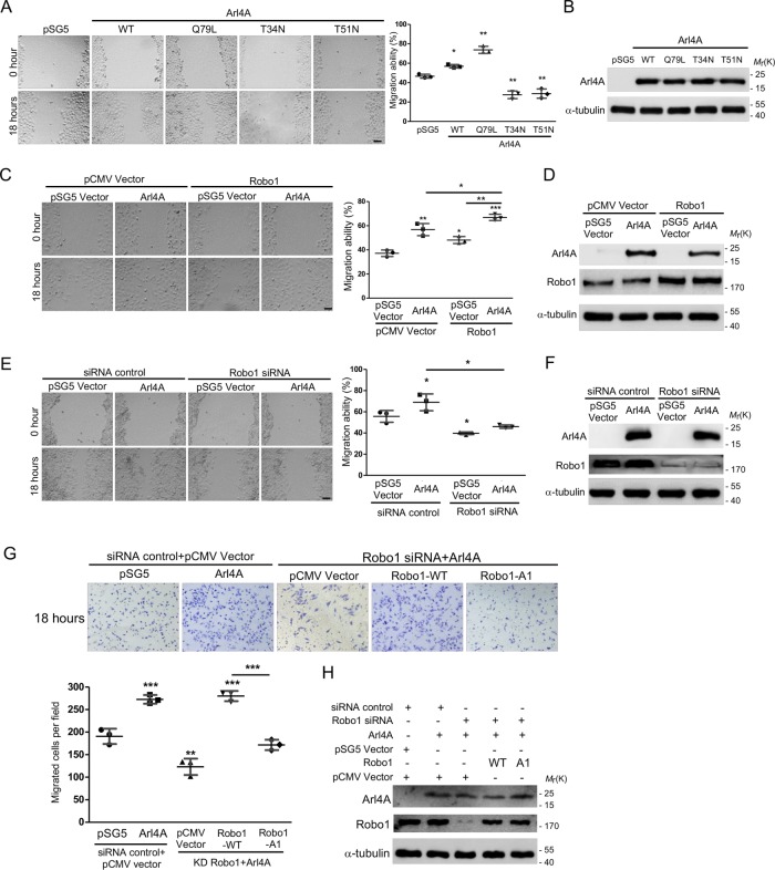
FIGURE 4:
The Arl4A-Robo1 interaction is necessary for increased cell migration. (A, C, E) Representative images of wound healing assays. HeLa cells were transfected with the indicated plasmids or siRNA for 18 h and then subjected to wound healing migration assays. Scale bar = 45 µm. Histograms: Wound healing migration data were quantified based on three biological replicates. (B, D, F) Total protein (20 μg) was loaded onto a 10-well gel to detect proteins. Western blot analysis of lysates from HeLa cells transfected with the indicated plasmids was performed to confirm equal expression. (F) The percentages of Robo1 after siRNA treatment were 23.1 ± 0.6% and 21.7 ± 0.4%. (G) Representative images of HEK293T cells transfected with the indicated plasmids and siRNA and then subjected to Transwell assays. The number of migrated cells in a field was calculated using ImageJ software after 18 h of migration. Histogram: Migration assay data were quantified based on three biological replicates. (H) Total protein (20 μg) was loaded onto a 10-well gel to detect protein expression. Immunoblotting analyses were used to evaluate protein expression levels in cells transfected with the indicated plasmids and siRNA. The percentage of Robo1 after siRNA treatment was 17.2 ± 0.5%. The solid bars represent the mean ± SD. *, P < 0.05; **, P < 0.005; ***, P < 0.001 (A: two-tailed Student's t test; C, E, and G: one-way ANOVA with Dunnett's post hoc multiple comparison test).

