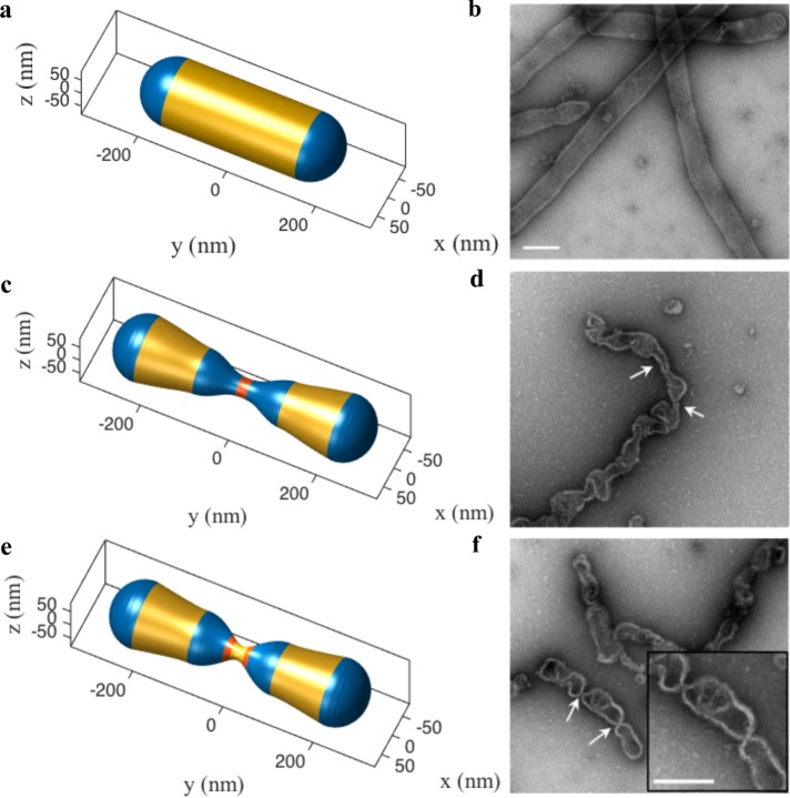
FIGURE 5:
In vitro validation of protein–lipid cooperativity during membrane squeezing. We simulate the constriction of a 150-nm-radius spherocylinder in the presence of the squeezing effect of Drp1 and conical lipids to compare to in vitro results presented in Stepanyants et al. (2015). As before, the spherocylinder undergoes extreme necking via buckling instability triggered jointly by curvatures from Drp1 and conical lipids. (a) The simulated geometry of an undeformed spherocylinder. (b) EM micrograph of the undeformed tubules (Stepanyants et al., 2015). (c) An intermediate simulated shape (Drp1 domain in red and high lipid concentration domain in blue). (d) EM micrograph of constricted tubules (Stepanyants et al., 2015). It is important to note that both c and d show similar local undulations and constrictions. (e) The postbuckling shape obtained by splitting of the Drp1 coat. (f) EM micrograph of tubules with highly constricted necks (Stepanyants et al., 2015). The narrow necks and the geometry of the neighboring domains show excellent agreement. Scale bar, 200 nm.

