
FIGURE 1:
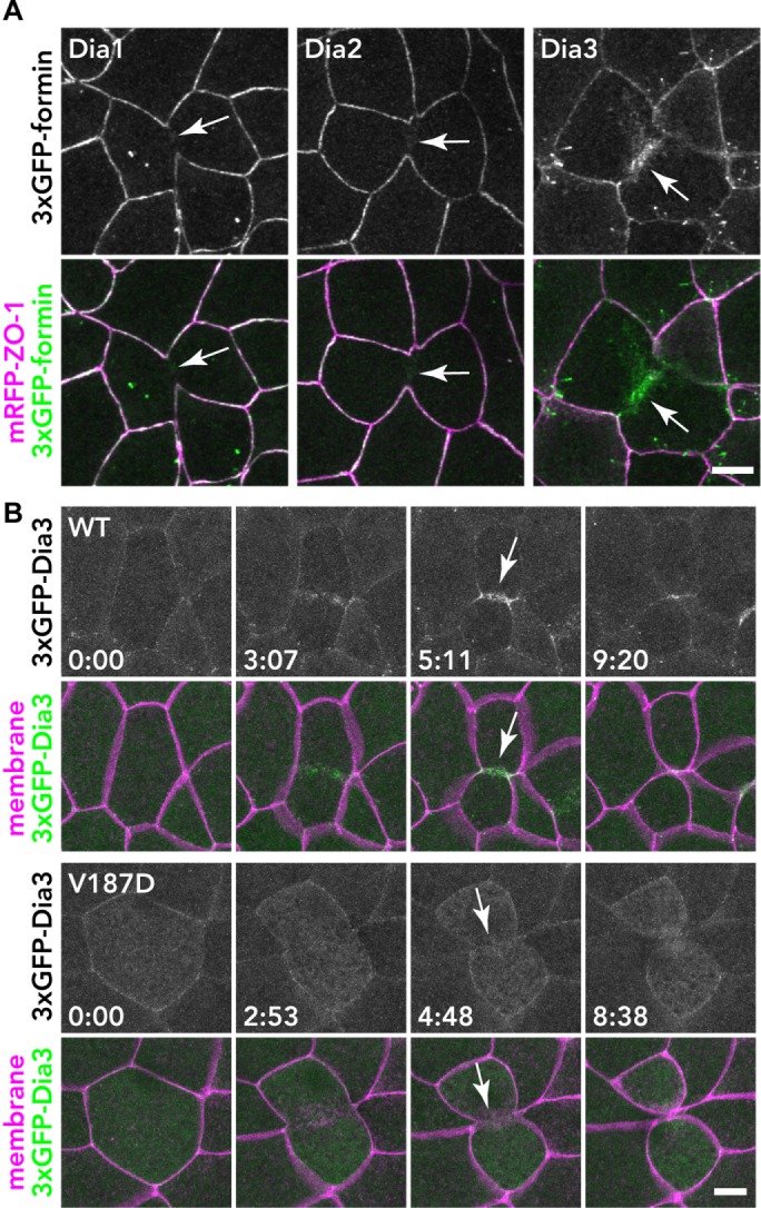
Localization of 3×GFP-tagged Dia1, Dia2, and Dia3 in the X. laevis gastrula epithelium. (A) Embryos expressing 3×GFP-tagged Dia1, Dia2, or Dia3 (green) and mRFP-ZO-1 (TJ marker; magenta) were live imaged using confocal microscopy; z-stack images of formin alone (top panels) and merged with mRFP-ZO-1 (bottom panels) are shown. Note that Dia3 is strongly localized at the contractile ring of the dividing cell. (B) The localization of Dia3 at the contractile ring is dependent on Rho binding. Embryos expressing 3×GFP-Dia1 WT or V187D (Rho-binding mutant; green) and mCherry-farnesyl (membrane probe; magenta) were imaged. Because the expression of Dia3 causes membrane deformation phenotypes (see A and Supplemental Figure S6), Dia3 was expressed at a lower level in these images. Note that the Dia3 V187D mutant cannot localize at the contractile ring. Scale bars: 10 µm.