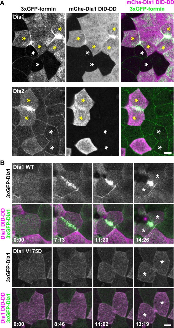
FIGURE 7:
Dia1 and Dia2 are localized at the contractile ring in dividing Dia1 DID-DD–overexpressing cells. (A) Embryos expressing 3×GFP-Dia1 (top panels) or 3×GFP-Dia2 (bottom panels) in all cells and mCherry-Dia1 DID-DD mosaically were live imaged using confocal microscopy. Note that Dia1 and Dia2 are strongly localized at the contractile ring in the Dia1 DID-DD–overexpressing cells (yellow asterisks) but are not localized at the contractile ring in the nonexpressing cells (white asterisks). (B) Embryos expressing 3×GFP-Dia1 WT (top panels) or 3×GFP-Dia1 V175D (bottom panels) in all cells and mCherry-Dia1 DID-DD mosaically were live imaged. Note that Dia1 V175D (Rho-binding mutant) cannot localize at the contractile ring in Dia1 DID-DD–overexpressing cells. Asterisks, daughter cells. Scale bars: 10 µm.

