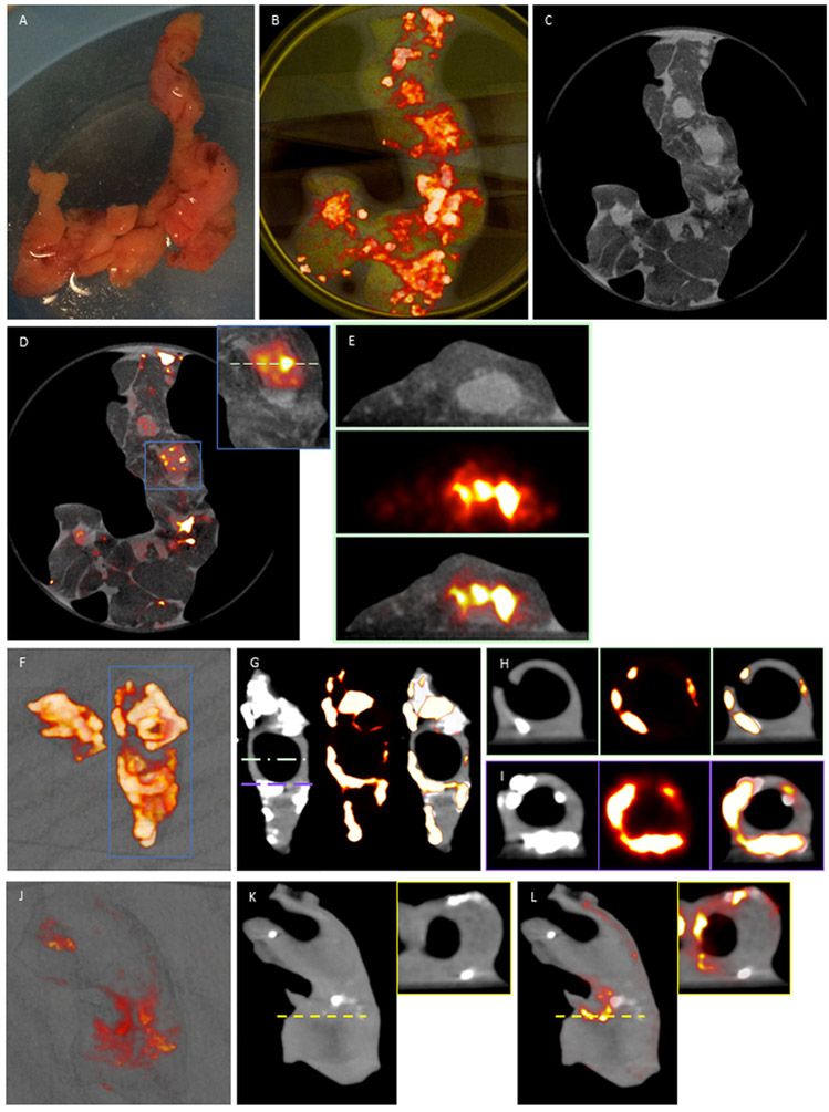
Figure 3. Coronary artery (a-e) and carotid endarterectomy specimens (f-l) 18F-fluoride μPET/CT.
A, Explanted coronary artery specimens were incubated in 100kBq/mL 18F-fluoride (t=60 mins). B, 3-dimensional volume rendered casts colocalizes binding to coronary artery sections with paucity of uptake in the surrounding epicardial fat and myocardium C, μCT and, D, fused images enabled, E, detailed axial reconstruction of 18F-fluoride binding in non-calcified coronary artery walls. F & J, 3-dimensional volume rendered casts of 18F-fluoride binding in explanted carotid artery specimens. G & K, sagittal CT colocalizes 18F-fluoride binding to exposed surfaces of hydroxyapatite on macrocalcified tissue. H, I, K (inset) & l (inset), focal 18F-fluoride binding is present in non-calcified regions of the carotid artery wall.

