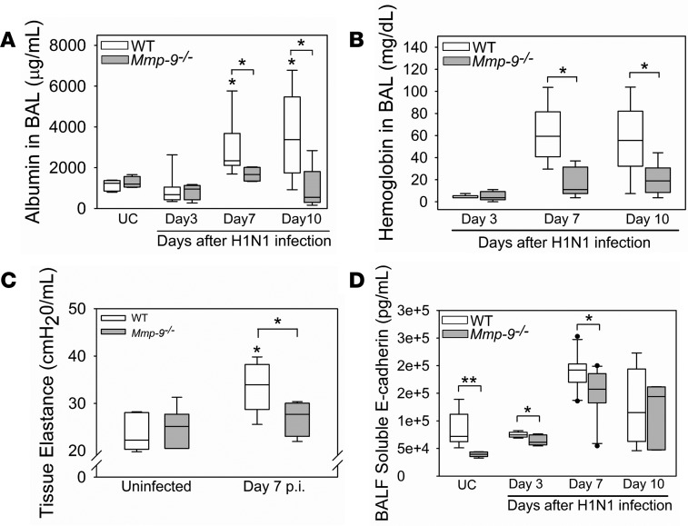
Figure 10. Mmp-9–/– mice were protected from lung injury following IAV infection.
WT and Mmp-9–/– mice were infected with an LD20 inoculum of H1N1 by the intranasal route. At postinfection intervals, albumin (A) and hemoglobin (B) levels were measured in BALF samples from H1N1-infected mice (4–11 mice/group) and in uninfected control (UC) mice (4–5 mice/group). *P < 0.05 versus the group indicated or uninfected controls belonging to the same genotype. (C) Tissue elastance was measured in H1N1-infected mice on day 7 p.i. (8 mice per group) and uninfected control mice (8–9 mice per group) using a FlexiVent device. *P < 0.05 versus the group indicated or UC mice belonging to the same genotype. (D) Soluble E-cadherin levels were measured in BALF samples from infected mice (4–11 mice per group) and uninfected control mice (6 mice per group) using an ELISA. *P < 0.05; **P < 0.01 versus the group indicated. All box-and-whisker plots show the medians and 25th and 75th percentiles, and the whiskers show the 10th and 90th percentiles. All data were analyzed with 1-way ANOVA followed by pair-wise testing with Mann-Whitney U tests.

