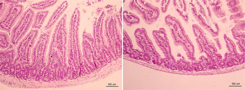
Fig. 2.

Representative images of the duodenal tissue of WT mice (n = 7) (left panel) and KO mice (n = 10) (right panel). Left: Villi adopt a long finger-like projection covered by a columnar epithelium. The core of lamina propria is colonized specially with many immunocompetent cells. Right: In KO mice, villi presented shorter and broader, with a normal epithelium
