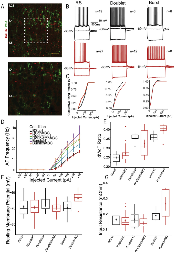
Fig. 3.
Impact of PNN digestion on putative excitatory neurons. (A) Example images of colocalization of WFA-labeled PNNs with SATB2, a marker of L5 pyramidal neurons in the barrel cortex. Scale bar represents 200 μM (top) and 100 μM (bottom). Representative traces of neurons classified as Regular Spiking (RS) Doublet and Burst neurons in control (top, black) and chABC digested sections (bottom, red). (C) Firing probabilities for RS, Doublet and Burst neurons. Putative excitatory neurons did not result in alterations to dV/dT ratio (E), resting membrane potential (F) or input resistance (G).

