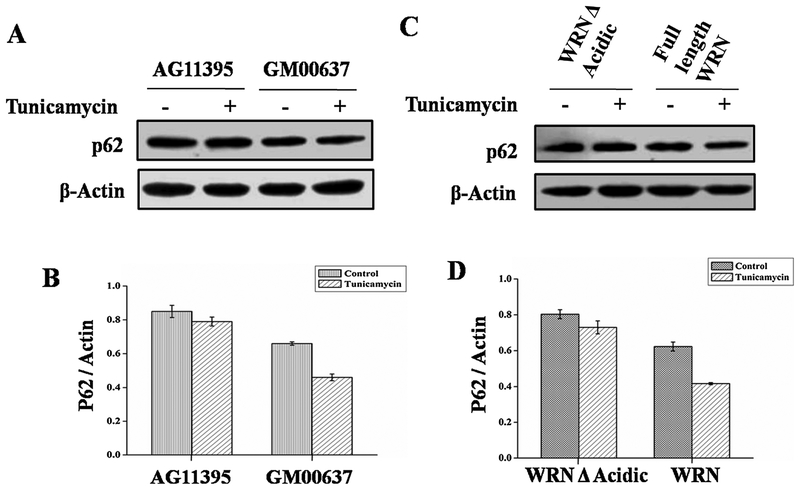
Fig. 5.
AG11395 and GM00637 cell lines were treated with ER stressor tunicamycin for 8 h along with complete medium. (A) Immunobloting of total cellular extract for autophagy flux marker protein p62. (B) Graphical representation of ratio of p62 and Actin. (C) AG11395 cell was transfected with wild type WRN and WRN Δ Acidic. Whole cell lysate was immunoblotted with anti-p62. (D) Graphical representation of p62 and Actin ratio. Here β-Actin was used as a loading control. 3 western blots from 3 separate sets of samples were used.

