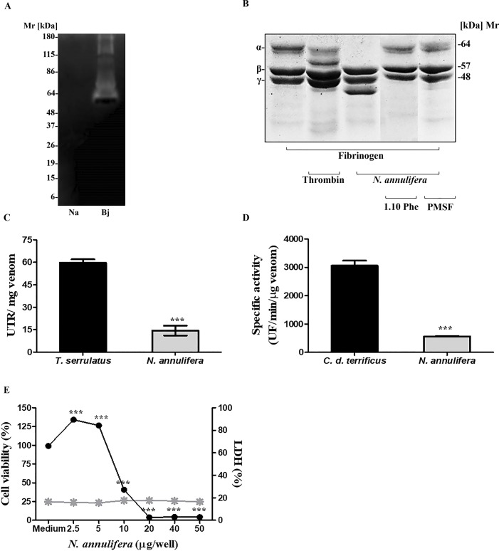
Fig 2. Toxic-enzyme properties of N. annulifera venom.
[A] Zymography: Samples of N. annulifera venom (30 μg) were assessed via 10% SDS-PAGE in the presence of 1 mg/mL gelatin and then incubated with the substrate buffer. Gels were stained with Comassie Brilliant Blue. The venom of B. jararaca (10 μg) was used as a positive control. [B] Fb cleavage: Fb samples (30 μg) were incubated with N. annulifera venom (5 μg) with or without metallo- (10 mM) and serine proteinase (10 mM) inhibitors for 1 hour. Samples were then analyzed via SDS-PAGE (8–16% gradient gel) and stained with Comassie Brilliant Blue. [C] Hyaluronidase activity: samples of N. annulifera venom (20 μg) were incubated at 37°C for 15 minutes in a solution containing hyaluronic acid. After the incubation, the reactions were stopped with CTAB and the absorbances were measured at λ 405 nm with a spectrophotometer. As a positive control, T. serrulatus (20 μg) scorpion venom was used. The results are representative of three separate experiments and expressed as UTR/mg of venom ± SD. Statistical analysis was performed using the t-test (*** p≤ 0.05). [D] PLA2 activity: Samples of N. annulifera venom (0.5 μg) were incubated at 37°C with a phospholipid mix that contained 10 mM phosphatidylcholine and 10 mM phosphatidylglycerol. Increased fluorescence was measured for 10 minutes. As a positive control, C. d. terrificus venom (0.5 μg) was used. The results are representative of three separate experiments and expressed as specific activity (UF per μg of venom per minute) ± SD. Statistical analysis was performed using t-test (*** p≤ 0.05). [E] Cytotoxic activity: The HaCat human keratinocyte cell lineage was cultured in DMEM medium and incubated during 72 hours with increasing amounts of N. annulifera venom. The effect on cell viability was evaluated using the MTT method and by measuring LDH release from human keratinocytes exposed to the venom, using the CytoTox 96 Non-Radioactive Cytotoxicity Assay Kit. Statistical analysis was performed using One Way ANOVA (*** p≤ 0.05) ± SD.

