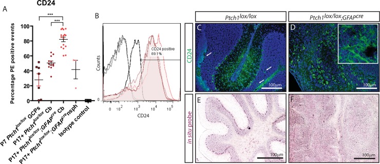
Fig 1. CD24 expression is elevated in P17+ primary Ptch1lox/lox;GFAPcre cerebellar cells compared to P7 granule cell precursors and P17+ wild type cerebellar cells.
(A) CD24 expression profiles of cerebella-isolated primary cells. (B) FACS histogram of CD24 expression on P17+ Ptch1lox/lox;GFAPcre medulloblastoma derived cells. (C-F) Immunofluorescent and in situ hybridisation stains for CD24 expression in murine models. CD24 expression was detected in the white matter, Purkinje cells and their dendritic extensions in P17+ Ptch1lox/lox cerebella as well as on random cells within the IGL (white arrows) following immunofluorescent analysis (C) and in situ hybridisations (E). In P17+ Ptch1lox/lox;GFAPcre medulloblastoma, CD24 expression was observed throughout the majority of the tumour cell mass (D + inset, F). Scale bars in C-F 100μm.

