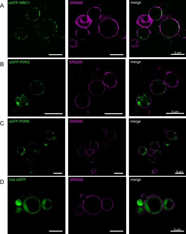
Fig 5. Confocal fluorescence microscopy of eGFP-PDR8, eGFP-PDR2 and eGFP-WBC1 in P. pastoris.
P. pastoris cells expressing either the fusion proteins eGFP-WBC1 (row A), eGFP-PDR2 (row B), or eGFP-PDR8 (row C) were analyzed by confocal fluorescence microscopy. As a control cells expressing free eGFP (row D) were analyzed as well. The left lane shows the eGFP fluorescence signal of the respective fusion protein, the middle lane the SR220 signal and the right lane the merged signals.

