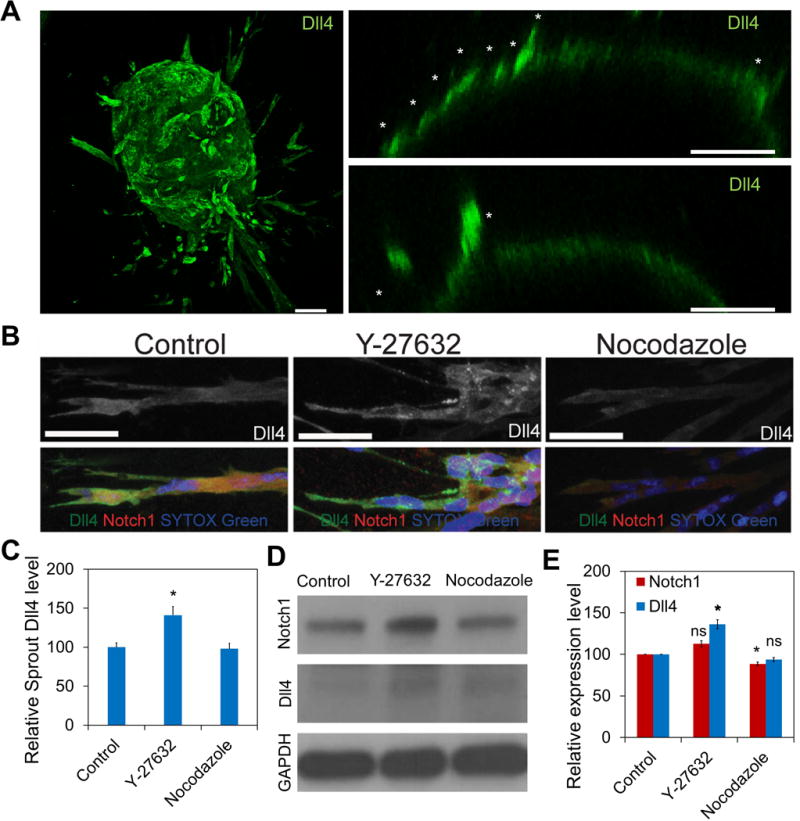
Figure 4. Mechanical force modulates Dll4 expression in sprouting embryoid bodies.

(A) Confocal image of a sprouting embryoid body (left). Sprouting embryoid bodies with Dll4-positive tip cells (white asterisks) near the surface of the embryoid body are indicated (right). Images are representative of five independent experiments. Scale bars, 100 μm. (B) Confocal images of Dll4 (red) and Notch1 (green) distributions in sprouts of embryoid bodies treated with Y-27632 and nocodazole. White asterisks indicate Dll4-positive cells in the sprout. Images are representative of three independent experiments. Scale bars, 50 μm. (C) Quantification of Dll4 expression in the sprout. (n = 6; ns, not significant; *P<0.05, **P<0.01, ***P<0.001; unpaired Student’s t-test). (D-E) Western blot analysis of Notch1 and Dll4 expression in embryoid bodies at day 5. Gene expression was normalized to GAPDH. Data are representative from three independent experiments and are expressed as mean ± s.e.m. (n = 3; ns, not significant; *P<0.05, **P<0.01, ***P<0.001; unpaired Student’s t-test).
