
Fig. 1.
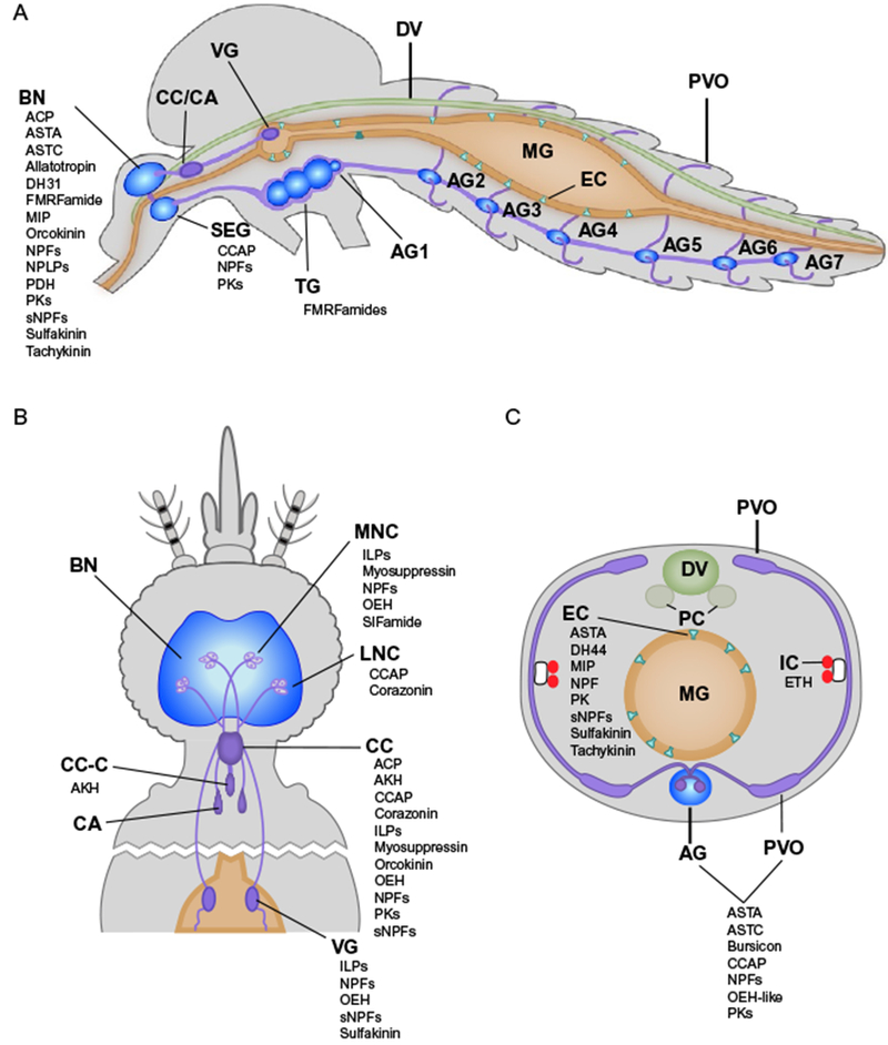
Schematic illustrating the sources of different peptide hormones in adult female mosquitoes as determined by immunocytochemistry or peptidomics. Sagittal view of the whole body (A), dorsal view of the head and thoracic regions (B), and a cross section through the third abdominal segment (C). Select regions of the nervous system, organs and cells are labelled in bold. Peptide hormones in Table 1 are listed alphabetically below detected sites. Abbreviations: AG1–7, abdominal ganglia 1 through 7; BN, brain; CA, corpora allata; CC, corpus cardiacum; CC-C, corpus cardiacum intrinsic cells; DV, dorsal vessel; EC, gut endocrine cells; IC, Inka cells; LNC, lateral neurosecretory cells; MG, midgut; MNC, medial neurosecretory cells; PC, pericardia; PVO, perivisceral organs; SEG, subesophegial ganglion; VG, ventricular ganglion. Peptide hormone abbreviations are listed in Table 1. Most of these data derive from studies of adults with the exception being for ETH, which is shown in (C) although data derive from studies of larvae (see text).