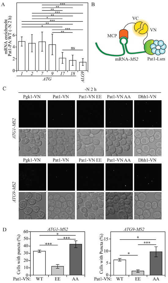
Fig. 4. Pat1 binds specific ATG mRNAs.
A) RNA immunoprecipitation was performed in Pat1-PA and WT (untagged) cells. Enrichment (Pat1-PA: WT) of ATG1, ATG2, ATG7, ATG9, ATG17, ATG18 and ALG9 (control) mRNA after 2 h of nitrogen starvation is presented. Error bars indicate the standard deviation of at least 5 independent experiments. ANOVA, * P<0.05, ** P<0.01, *** P<0.001, ns, no statistical significance B) Schematic model for protein-RNA BiFC. Interaction between MS2 coat protein (MCP) tagged with C-terminal vYFP (VC) bound to an MS2 hairpin-tagged ATG mRNA and Pat1-tagged with N-terminal vYFP (VN), leads to a fluorescent signal by a complete vYFP protein. C) Protein-RNA BiFC was used to determine the interaction of Pgk1-VN, Pat1-VN, Pat1-VN AA, Pat1-VN EE and Dhh1-VN with ATG1-MS2- and ATG9-MS2-tagged mRNA after 2 h of nitrogen starvation (−N). D) Quantification of the number of cells showing puncta in Pat1-VN, Pat1-VN EE and Pat1-VN AA cells also expressing either ATG1-MS2- or ATG9-MS2-tagged mRNA. Error bars indicate the standard deviation of at least 3 independent experiments. ANOVA, *P <0.05 and *** P <0.001. See also Figure S4.

