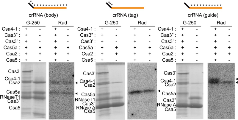
Fig. 8.

Identification of direct interactions between crRNA and Csa proteins by UV crosslinking. SDS-polyacryladmide gel showing Coomassie blue (G-250) stained Csa proteins and RNase T1 and/or RNase A in each reaction and corresponding autoradiograph images (rad) showing crRNA-interacting Csa proteins (black triangles).Reactions were incubated with either 2 or 6 Csa proteins (same as Fig. 7b, c). The schematic above each panel indicates 32P and 4-thiouridine-labeled crRNAs used in each experiment (5′ tag sequence (black), guide sequence (orange) and radiolabeled regions (asterisks)
