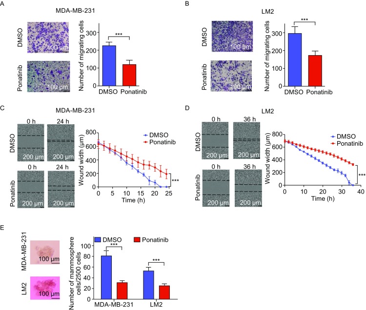
Figure 2.

Ponatinib inhibits breast cancer migration in vitro. (A and B) The effect of Ponatinib on the migration of MDA-MB-231 cells (A) and LM2 cells (B) was tested by a Transwell migration assay. Cells were treated with 1 µmol/L Ponatinib for 8 h, the migrating cells were stained with crystal violet (left panel) and quantified (right panel). ***P < 0.001 by two-sided Student’s t-test. Scale bar, 100 µm. (C and D) The effect of Ponatinib on the migration of MDA-MB-231 cells (C) and LM2 cells (D) was tested by a wound healing assay. Cells were treated with 1 µmol/L Ponatinib, the wound width was photographed after scratching (left panel) and quantified (right panel). P value by one-tailed Mann-Whitney U test. Scale bar, 200 µm. (E) The effect of Ponatinib on mammosphere formation of MDA-MB-231 cells and LM2 cells was tested by an ultralow attachment microplate. Cells were treated with 1 µmol/L Ponatinib. ***P < 0.001 by two-sided Student’s t-test. Scale bars, 100 µm. Representative images of one of three independent experiments are shown. Error bars represent mean ± SD of 6 samples in each group (A–E)
