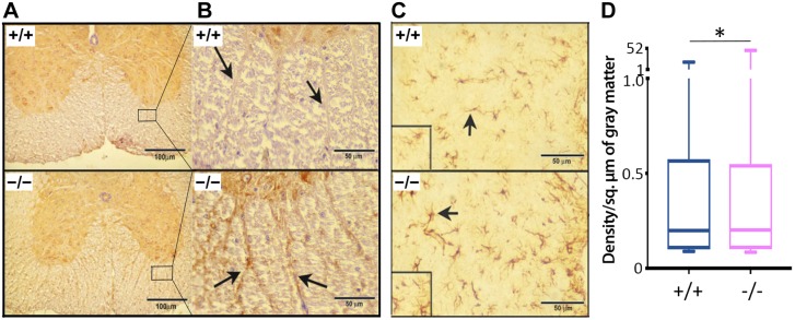
Figure 5 .
Immunohistochemical staining for myelin basic protein in a cross section of the SC. A, B) The overall increase in the expression of MBP can be seen in white matter (anterior funiculus shown in the magnification) with intense staining along the tracts (arrows) in the Cd320−/− mice. C) Increased expression of GFAP in the SC of Cd320−/− mice (arrows; intermediolateral gray column). D) Quantification of immunostaining for GFAP (n = 6). *P < 0.05.

