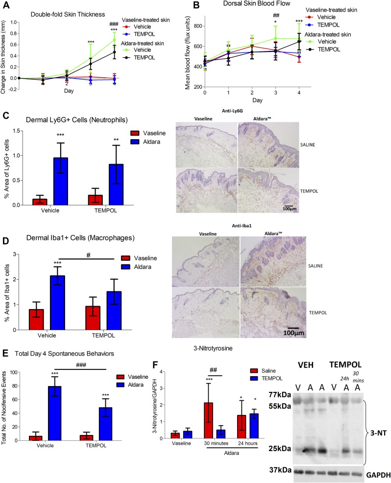
Figure 6 .
Tempol protected against Aldara-mediated skin pathology. A) Changes in double-fold skin thickness (mm). B) Dorsal skin mean blood flow (flux units). C) Grouped data of percentage of area of dermal Ly6G+ cells using automated analysis and representative images of skin sections (right). Hematoxylin counterstaining. D) Grouped data of percentage of area of dermal Iba1+ cells and representative images of skin sections (right). Hematoxylin counterstaining. E) Total d 4 spontaneous behaviors. F) Grouped data for 3-NT and representative blot for 3-NT and GAPDH (right). Graphs represent means ± sd and were analyzed by 2-way or repeated measures ANOVA with Bonferroni’s post hoc test; n = 4–7 animals/group. Original maginification, ×10. Scale bars, 100 μm. *P < 0.05, **P < 0.01, ***P < 0.001 between Vaseline vs. Aldara groups, #P < 0.05, ##P < 0.01, ###P < 0.001 between VEH vs. tempol. A, Aldara, V, Vaseline.

