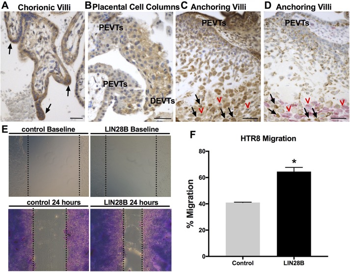
Figure 3 .
LIN28B is highly expressed in syncytial sprouts and invasive trophoblasts in first trimester placentas and increases trophoblast migration in vitro. A–C) Representative images of immunohistochemical staining for LIN28B in human first trimester placenta. LIN28B is highly expressed in syncytial sprouts of chorionic villi (black arrows, A). LIN28B expression increases from proximal EVTs (PEVTs) to distal invasive EVTs (DEVTs) in placental cell columns (B). LIN28B expression is lowest in PEVTs and increased in interstitial trophoblasts (black arrows) invading maternal decidua (red arrowheads) in anchoring villi (C). D) Representative serial section of tissue shown in C double immunostained for DC marker vimentin (pink) and trophoblast marker cytokeratin-7 (brown). Red arrowheads and black arrows in D correspond to red arrowheads and black arrows in C. E) Representative results of scratch wound assay at time of scratch application (top) and after 24 h (bottom) from HTR8/SVneo cells transfected with either GFP (control) or LIN28B. F) Quantification of migration after 24 h. Scale bars, 35 mm. Results represent means ± sem. *P < 0.05 vs. control.

