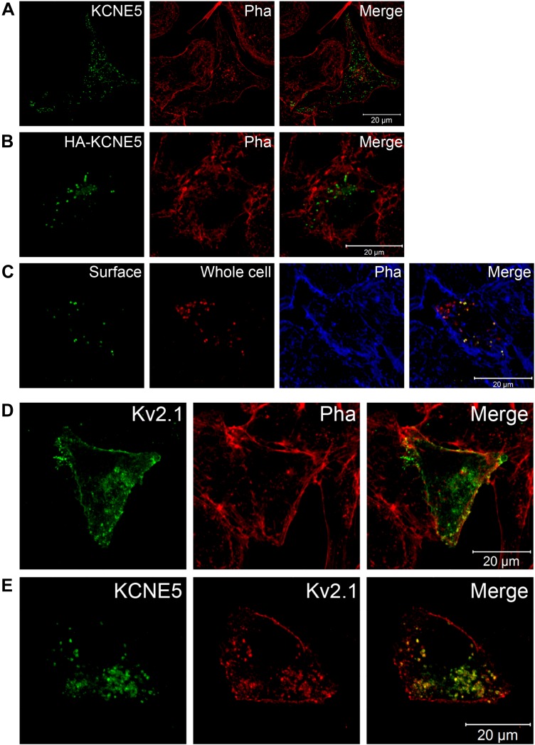
Figure 6 .
KCNE5 and KV2.1 colocalize in HL-1 cells. A, B) Confocal images of HL-1 cells heterologously expressing WT KCNE5 (A) or HA-tagged WT KCNE5 (B). Phalloidin (Pha) labeled the submembranous actin cytoskeleton and was used to visualize the cell membrane. C) Nonpermeabilized HA-KCNE5–expressing HL-1 cells stained with a specific HA-tagged antibody (surface staining, green), permeabilized, and then stained with a KCNE5-specific antibody (red) and Phalloidin (blue). D) Colabeling of single transfected KV2.1 (green) and endogenous Phalloidin (red) in HL-1 cells. E) Colabeling of cotransfected KCNE5 (green) and KV2.1 (red) in HL-1 cells.

