Figure 2 .
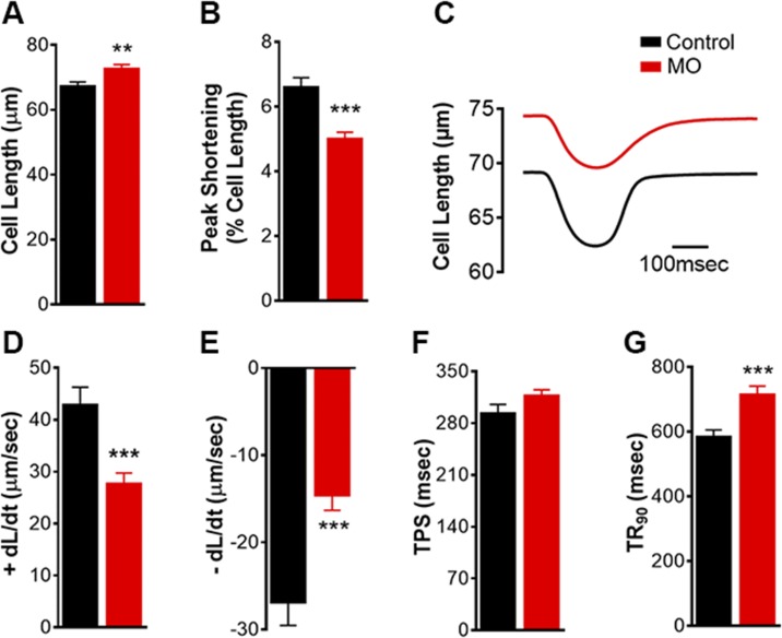
Mechanical contractile properties based on cell-length measurement of LV cardiomyocytes from 0.9 gestation fetuses of control and MO mothers. Resting cell length (A); PS (B); representative contractile trace (C); maximum velocity of shortening (D); maximum velocity of relengthening (E); TPS (F); and TR90 (G). Means ± sem (n = 140 cells: 13–33 cells/animal from 5 control fetal hearts; n = 100 cells: 11–32 cells/animal from 7 MO fetal hearts). **P < 0.01, ***P < 0.001 vs. control.

