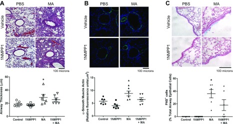
Figure 3 .
Histology of airway inflammation in TrkBKI mice. H&E and PAS stained lung sections show airway inflammation, α-smooth muscle actin expression, and mucus production in MA-challenged mice. Airway inflammation and thickness (A), α-smooth muscle actin expression (B), and mucin expression (C) were not significantly reduced in mice administered 1NMPP1. Original magnification: ×200 (A, B), ×400 (C). Data are presented as means ± sem (n = 6 mice/group). *P < 0.05, significant difference between control and MA.

