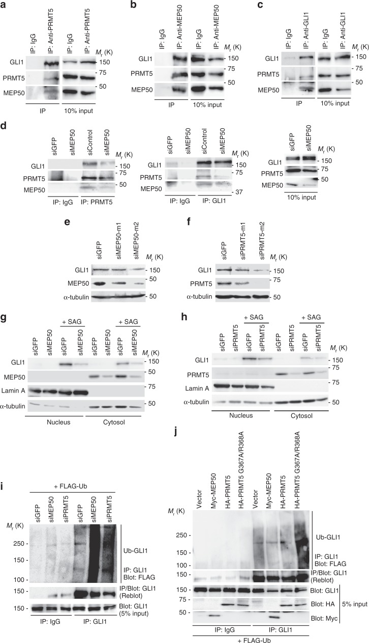
Fig. 2.
MEP50/PRMT5 complex supports GLI1 activation through GLI1 stabilisation downstream of the HH signalling pathway. a–c Endogenous GLI1/MEP50/PRMT5 complex in C3H10T1/2 cells. Cells were treated with SAG for 36 h, and complex was detected by immunoprecipitation (IP) with anti-PRMT5 (D5P2T) (a), anti-MEP50 (ERP10708 [B]) (b), or anti-GLI1 (V812) (c) antibodies, followed by immunoblot (IB) with antibodies against indicated proteins. d Dissociation of PRMT5 and GLI1 in stable MEP50 knockdown C3H10T1/2 cells by siMEP50-m2. Cells were treated with SAG for 48 h and treated with 50 μM MG132 for 4 h. GLI1/PRMT5 complex was detected by immunoprecipitation with anti-PRMT5 (D5P2T) or anti-GLI1 (V812) antibodies, followed by immunoblot with indicated antibodies. e Immunoblot of endogenous GLI1 in C3H10T1/2 cells expressing MEP50 siRNAs (siMEP50-m1 or siMEP50-m2). f Immunoblot of endogenous GLI1 in C3H10T1/2 cells expressing two independent PRMT5 siRNAs. g Immunoblot of nuclear and cytoplasmic GLI1 and MEP50 in stable MEP50-knockdown (siMEP50-m2) or control siGFP-expressing cells treated with 300 nM SAG. Cells were treated with SAG for 24 h and separated into cytosol and nucleus fractions. h Immunoblot analysis of endogenous nuclear and cytoplasmic GLI1 and PRMT5 in stable PRMT5-knockdown (siPRMT5-m2) C3H10T1/2 cells. Cells were treated with 300 nM SAG for 24 h and then separated as in h. i In vivo ubiquitination of GLI1 in C3H10T1/2 cells with or without expression of siMEP50. FLAG-ubiquitin was transfected into C3H10T1/2 cells. After 48 h of transfection, then cells were treated with 50 µM MG132 for 4 h. Endogenous ubiquitinated GLI1 was immunoprecipitated with an anti-GLI1 (C-1) antibody, followed by immunoblotting with indicated antibodies. j In vivo ubiquitination of GLI1 in C3H10T1/2 cells with exogenous expression of PRMT5 or MEP50. FLAG-ubiquitin and HA-PRMT5, HA-PRMT5 G367A/R368A (inactive form of PRMT5), or Myc-MEP50 were transfected into C3H10T1/2 cells. After 48 h, the cells were treated with 50 µM MG132 for 4 h. Endogenous ubiquitinated GLI1 was detected as described in i. In a, i and j, data represent one of two independent experiments with similar results. Unprocessed original scans of blots are shown in Supplementary Fig. 6

