Fig. 5.
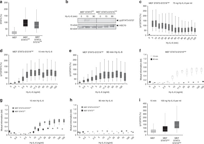
STAT3-S727 phosphorylation increases robustness of STAT3 phosphorylation against varying STAT expression. a STAT3 expression in MEF, MEF STAT3high, and MEF STAT3-S727Ahigh cells was evaluated by flow cytometry using fluorescent antibodies against STAT3. For independent experiments mean fluorescence of cells per cell line was calculated. Mean fluorescence of STAT3 expression in MEF STAT3high cells was set to 100%. The data are from n = 3 experiments. b MEF STAT3high and MEF STAT3-S727Ahigh cells were stimulated with 75 ng Hy-IL-6 per ml for the indicated times. STAT3-S727 phosphorylation and HSC70 expression were evaluated by Western blotting. A representative result of n = 6 independent experiments is shown. Uncropped Western Blots are shown in Supplementary Figure 12a, b. c MEF STAT3-S727Ahigh cells were stimulated with 75 ng Hy-IL-6 per ml for the indicated times. STAT3 phosphorylation was evaluated by flow cytometry. d MEF STAT3-S727Ahigh cells were stimulated with increasing amount of Hy-IL-6 for 15 min. STAT3 phosphorylation and expression were evaluated by flow cytometry using fluorescent antibodies against STAT3 (pY)705 and STAT3 (Supplementary Figure 13). The data are pooled from n = 3 experiments. e MEF STAT3-S727Ahigh cells were stimulated with increasing amount of Hy-IL-6 for 90 min and analysed as in d) (Supplementary Figure 14). For independent experiments, mean fluorescence of cells per cytokine dose was calculated. Maximal mean fluorescence in each experiment was normalised to 100 %. Data are from n = 3 experiments. f Based on data presented in Fig. 5d and Supplementary Figure 13, or Fig. 5e and Supplementary Figure 14, Mutual Information between STAT3 expression and IL-6-induced STAT3 phosphorylation in MEF STAT3-S727Ahigh cells stimulated with Hy-IL-6 for 15 min (white) or 90 min (black) was calculated. Data are from n = 3 independent experiments. g Overlay of Fig. 3d and Fig. 3f. h Overlay of Fig. 4f and Fig. 5f. i MEF, MEF STAT3high, and MEF STAT3-S727Ahigh cells were stimulated with 100 ng Hy-IL-6 per ml for 15 min. STAT3 phosphorylation in MEF, MEF STAT3high, and MEF STAT3-S727Ahigh cells was evaluated by flow cytometry. For independent experiments mean fluorescence of cells per cell line was calculated. Mean fluorescence of STAT3 phosphorylation in MEF STAT3high cells was set to 100%. The data are from n = 3 experiments

