Fig. 1.
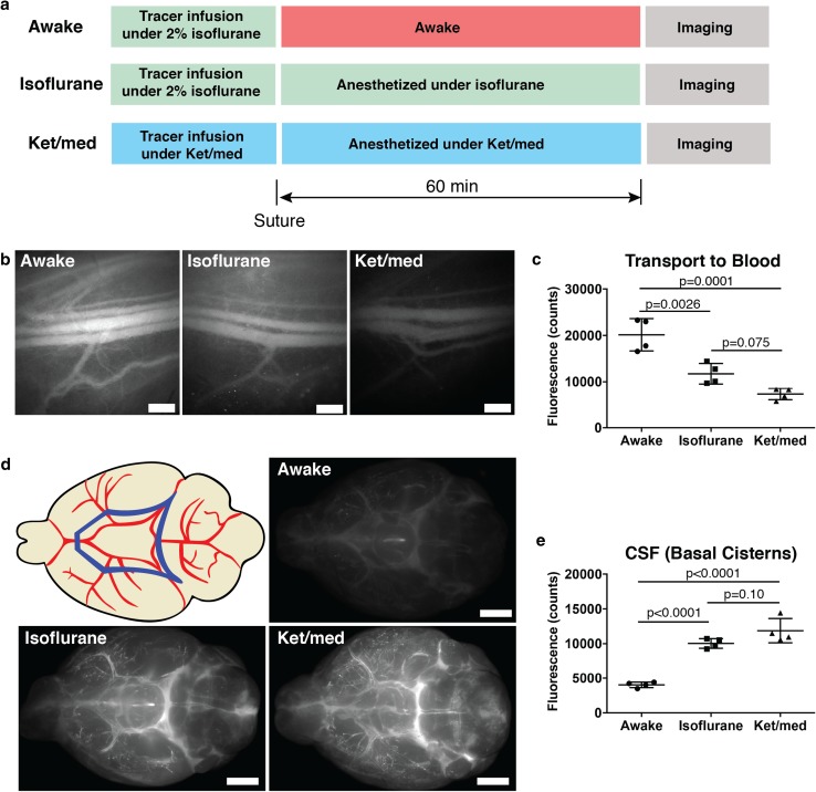
CSF outflow is increased during awake conditions. a Scheme of experimental design for quantification of CSF outflow during awake or anesthesia conditions. Before imaging at the saphenous vein, mice from awake and isoflurane group were given 80 mg/kg ketamine, 0.2 mg/kg medetomidine intraperitoneally. After imaging, all mice were overdosed with 400 mg/kg ketamine, 1 mg/kg medetomidine. b Representative images of saphenous bundle of blood vessels at 60 min post-infusion of P40D680 into lateral ventricle. Scale bar: 500 µm. c Quantification of P40D680 signal in the blood 60 min after tracer i.c.v. infusion. d Scheme indicating ROI and ex vivo imaging of the basal aspect of the brain at 60 min post-infusion showing presence of P40D680 tracer at circle of Willis and cisterns. Blue polygon in upper left image represents the analyzed ROI. Scale bar: 2000 µm. e Quantification of P40D680 signal at basal cisterns 60 min after infusion

