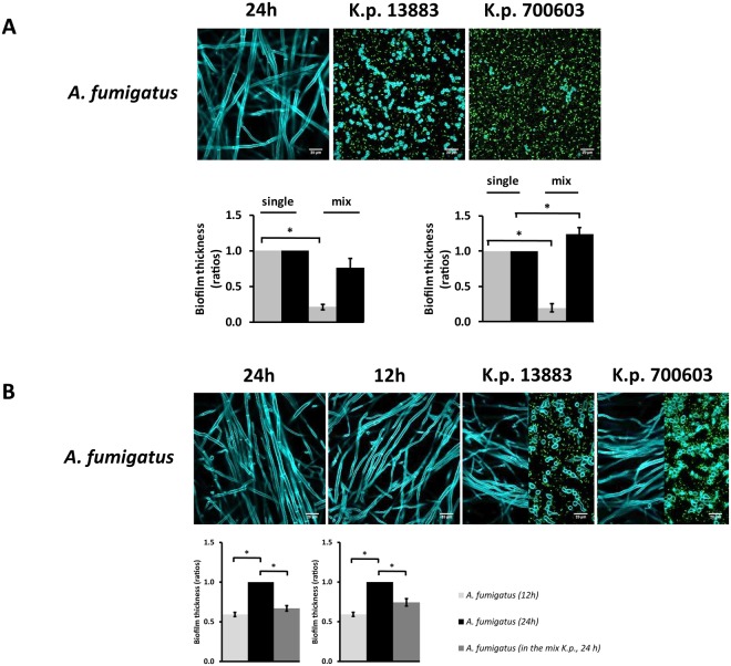
Figure 2.
Biofilm thickness of A. fumigatus vs K. pneumoniae analyzed by confocal microscopy. The fluorescent photos represent A. fumigatus growing alone or in co-culture with different strains of K. pneumoniae. Differential staining of the microorganisms upon growth in culture and measurement of biofilm thickness was performed as indicated in the Methods section. (A) Fungi and bacteria co-cultured since time zero. Grey bars correspond to fungi and black bars correspond to bacteria. (B) Fungal spores germinated for up to 12 h followed by addition of bacteria for a total of 24 h. The confocal microscopy images of A. fumigatus and K. pneumoniae co-cultures (K.p.13883, K.p.700603) show the levels of hyphae (left) and spores (right), respectively. The left plots indicate co-cultures of A. fumigatus with K. pneumoniae ATCC 13883 and the right plots indicate co-cultures of A. fumigatus with K. pneumoniae ATCC 700603. Biofilm thickness of the single cultures was set to one and the biofilm thickness of each species in the co-culture was normalized to the corresponding single culture. Data are presented as mean + SE of three independent experiments.

