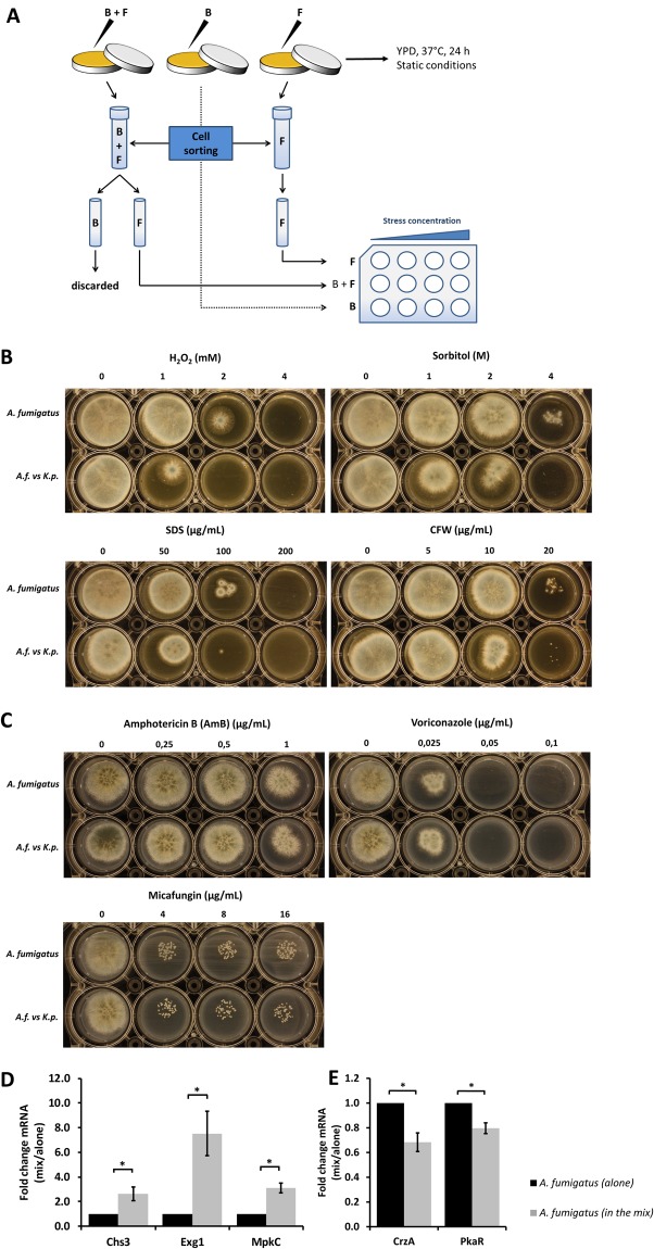
Figure 6.
Induction of cell wall stress and upregulation of cell wall-related genes in A. fumigatus mediated by K. pneumoniae. (A) Schematic view of flow sorting of A. fumigatus and K. pneumoniae (ATCC 700603). Sorted cells were plated onto 12-well agar plates containing kanamycin (1000 µg/mL) and colistin (100 µg/mL) for selection of fungal growth and prevention of any residual bacterial contamination. Plates also contained different concentration ranges of cell wall stressors and antifungal drugs to facilitate the observation of the sensitivity of fungi previously grown alone or exposed to K. pneumoniae. (B) Sensitivity of A. fumigatus to oxidative (H2O2), osmotic (sorbitol, SDS) and cell wall (CFW) stresses, with and without previous exposure to K. pneumoniae. (C) Sensitivity of A. fumigatus to antifungal drugs (amphothericin B, voriconazole, micafungin) with and without previous exposure to K. pneumoniae. (D) Upregulation of A. fumigatus cell wall-related genes in the presence of K. pneumoniae. (E) Downregulation of hyphae-related genes of A. fumigatus in the presence of K. pneumoniae determined by quantification of mRNA expression of hyphae-related genes of A. fumigatus upon exposure to K. pneumoniae (ATCC 700603). Fungal spores were germinated for up to 12 h followed by addition of bacteria for a total of 24 h. Transcript levels of the A. fumigatus single culture were set to one, and the transcript levels in the co-culture were normalized to the single culture. Additionally, normalization was also performed against the reference gene TUBA. Data are presented as mean + SE of four independent experiments.

