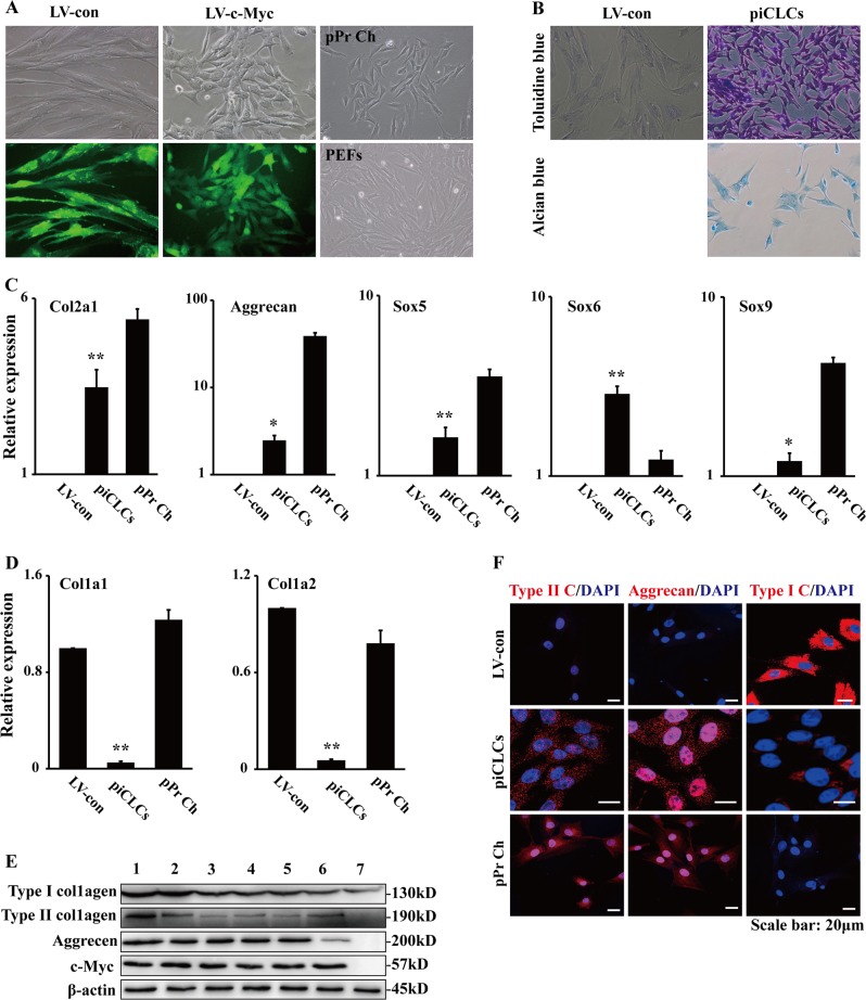
Fig. 3. Chondrocyte marker gene expression analyses of pig induced chondrocyte-like cells (piCLCs) from PEFs by c-Myc.
a Cell morphologies of PEFs, pPr Ch and piCLCs. The phase-contrast photographs of c-Myc-expressing PEFs (i.e., piCLCs) and vector-expressing PEFs were token at day 4 post-infection. pPr Ch: porcine primary chondrocytes. b Toluidine blue staining and alcian blue staining for piCLCs. Proteoglycan was identified by toluidine blue staining and alcian blue staining. c, d qRT-PCR analysis of chondrocyte (c) and fibroblast (d) marker gene expression in the indicated cells. In (c) and (d), error bars indicate mean ± SD (n = 3). e Cell extracts from pPr Ch, piCLCs and PEFs were analysed by immunoblotting with antibodies against the indicated proteins. Lane 1: pPr Ch; Lane 2: piCLCs P7; Lane 3: piCLCs P14; Lane 4: piCLCs P19; Lane 5: piCLCs P25; Lane 6: piCLCs-S; Lane 7: PEFs. f Immunofluorescence analysis showing protein expression of type I collagen (type I C), type II collagen (type II C) and aggrecan in the indicated cells. Nuclei were counterstained with DAPI

