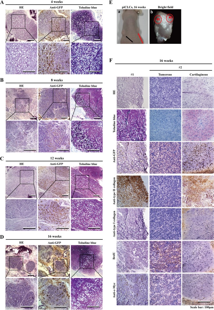
Fig. 7. Prolonged of in vivo cartilage formation in nude mice.
a–d Histology of grafted tissues 4 weeks (a), 8 weeks (b), 12 weeks (c), or 16 weeks (d) after injection of piCLCs (3 × 10⁶cells/site) into nude mice. Semiserial sections were stained with HE, toluidine blue and immunostained with anti-GFP antibody. For each tissue, lower-magnification images are shown above, and higher magnification images of boxed regions in the top panels are shown below. e Injection of piCLCs produced tumours (red arrow; e-a) at injected sites (16 weeks after injection). e-a Picture of nude mouse harbouring xenografts (arrows). e-b Picture of the xenograft (shown by red arrow in e-a) stripped from a nude mouse. Supplementary Fig. S6 and f show the histology of cartilage tissue and tumour tissue generated by piCLCs. f Semiserial sections of typical cartilage tissue (left column), tumorous portion (middle column) and cartilaginous portion (right column) formed from the transplanted piCLCs after injection were stained with HE and toluidine blue and immunostained with the indicated antibodies, as shown on the left. The typical cartilage tissue (left column) and the tumorous portion (middle column) and cartilaginous portion (right column) correspond to circled regions #1 and #2 in e-b, respectively

