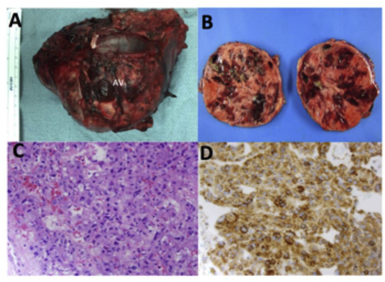
Fig. 2. Gross pathology and histopathology of large pheochromocytoma.
(A) Gross image of left adrenal mass resected with a pronounced left adrenal vein (AV). (B) Gross image of a bisected left adrenal mass showed variegated, partially cystic and hemorrhagic cut surface. (C) H&E staining of the adrenomedullary tumor cells showing finely granular eosinophilic cytoplasm, round to oval nuclei with occasional prominent nucleolus and focal cytologic pleo-morphism characteristic of pheochromocytoma. Magnification, 400 ×. (D) Immunohistochemical staining of the left adrenal tumor reveals retained succinyl dehydrogenase B (SDH-B) expression (brown staining) in the tumor cells. Magnification, 400 ×.

