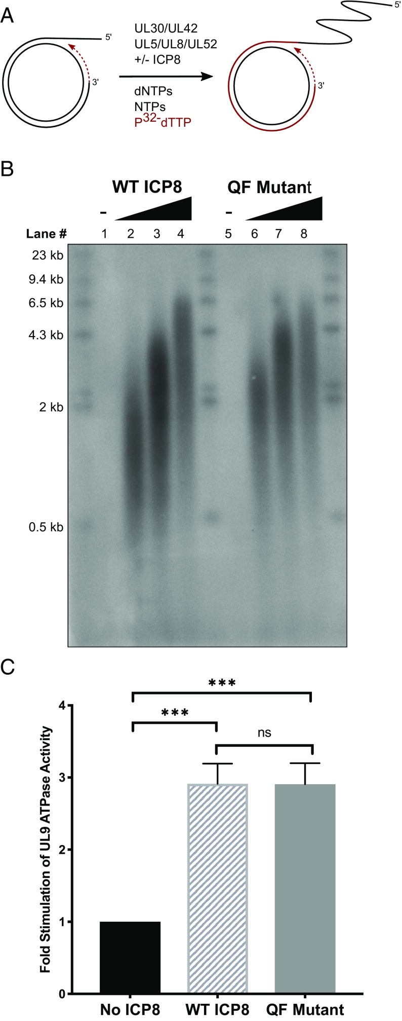
Fig. 4.
The QF mutant stimulates replication proteins in vitro. (A) Schematic of rolling-circle replication on a minicircle substrate comprised of a 70-mer ssDNA circle annealed to a 90-mer linear oligo. The 3′ end of the 90-mer acts as the primer for leading-strand synthesis. Leading-strand DNA synthesis is detected by incorporation of [α-32P]dTTP. Newly synthesized DNA is shown in red. (B) An alkaline agarose gel showing stimulation of in vitro leading-strand DNA synthesis by WT and QF mutant proteins. The minicircle assay was performed using increasing concentrations of either WT ICP8 or mutant QF protein (0, 190, 380, and 760 nM). After 60-min incubation at 37 °C, reactions were quenched, and replication products were separated by 1% alkaline agarose gel electrophoresis and imaged with a phosphorimager. (C) ICP8 stimulation of UL9 ssDNA-dependent ATPase activity. WT or QF mutant proteins (700 nM) were mixed with 100 nM UL9 in the presence of 5 mM ATP and 10 µM ssDNA. Reactions were incubated for 1 h at room temperature and quenched with EDTA. ATPase activity was measured by the release of free phosphate as described in Materials and Methods. Fold stimulation relative to the control lacking ICP8 is plotted on the graph. Error bars represent SD. P values were determined by ordinary one-way ANOVA with Tukey’s multiple-comparisons test (***P = 0.0001 and nsP = 0.9993).

