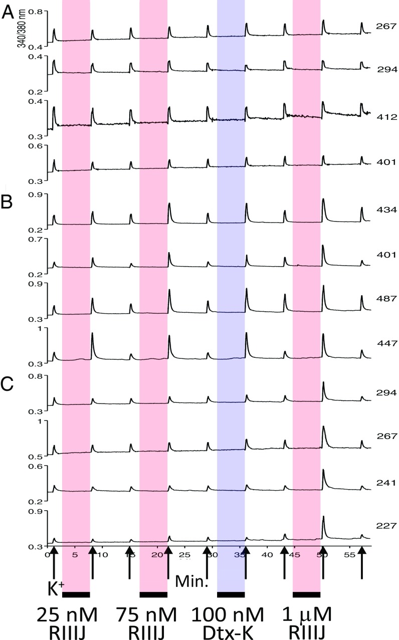
Fig. 4.
κM-RIIIJ can distinguish between different sensory neuron subtypes. Representative calcium-imaging traces from three DRG neuronal subclasses displaying distinct responses to κM-RIIIJ and Dtx-K. (A) Neurons unaffected by κM-RIIIJ or Dtx-K: Four representative neurons unaltered by the application of κM-RIIIJ (25 nM, 75 nM, and 1 μM) and Dtx-K (100 nM), suggesting the lack of functional expression of Kv1.1 and Kv1.2 channels in these neurons. (B) Neurons responsive to the application of low [κM-RIIIJ]. The examples shown correspond to neurons that displayed an increase in peak height following a depolarizing stimulus in the presence of 25 nM, 75 nM, and 1 μM κM-RIIIJ and 100 nM Dtx-K, indicating the presence of both Kv1.1 and Kv1.2 channels in these neurons. (C) Neurons exclusively responsive to high [κM-RIIIJ]. These neurons were not affected by the application of low doses (25 and 75 nM) of κM-RIIIJ nor 100 nM Dtx-K but displayed increases in peak height after exposure to 1 μM κM-RIIIJ. The low sensitivity to κM-RIIIJ and insensitivity to Dtx-K suggest the presence of homotetrameric Kv1.2 channels and the absence of homomeric Kv1.1 or heteromeric Kv1.1/1.2 complexes in this cell population. Each trace corresponds to individual cells. The x axis is time (in minutes), and the y axis tracks the ratio of relative fluorescence change (ΔF/F) excited at 340 and 380 nM. Upward arrows mark the application of the depolarizing stimulus (20 mM [K+]o, ∼15 s). Colored boxes (red: κM-RIIIJ; blue: dendrotoxin-K) indicate the time of peptide exposure (∼6 min) in between the applications of depolarizing stimulus. Numbers to the right indicate cell area in μm2.

