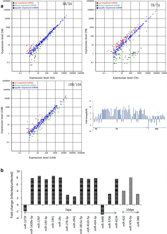
Fig. 1.
a Scatter plots and fold changes comparing the midguts of infected and uninfected Ae. albopictus at different time points. 5B/5A is the (5-day uninfected midguts)/(5-day infected midguts after exposure to DENV-2), 7B/7A is the (7-day uninfected midguts)/(7-day infected midguts), etc. b Screened significantly differentially expressed miRNAs between the midguts of infected and uninfected Ae. albopictus at different time points after a DENV-2-infected blood meal

