Fig. 1.

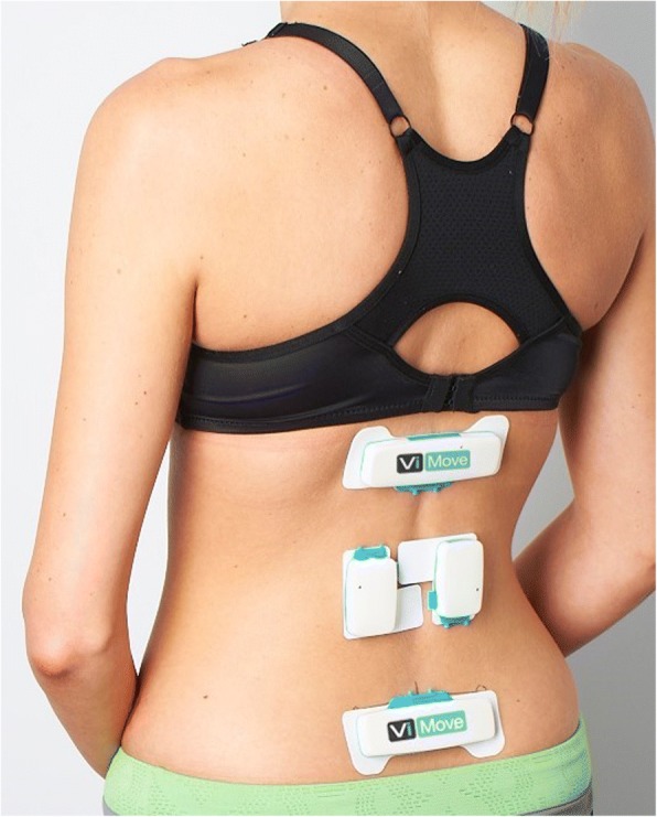
Device Placement. An example of sensor placement with the lower border of the upper sensor placed at the T12 level, the upper border of the lower sensor level with S1 and the EMG sensors placed over lumbar extensor muscles at the level of L3

Device Placement. An example of sensor placement with the lower border of the upper sensor placed at the T12 level, the upper border of the lower sensor level with S1 and the EMG sensors placed over lumbar extensor muscles at the level of L3