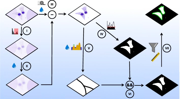
Scheme 1.
Steps in image processing and segmentation algorithm. (I,II): Production of a subtraction mask for background subtraction by duplicating the raw image and constraining maximum to three-fold the mean gray value of the image (I). Gaussian blurring (shown here as the water droplet) of the constrained image (II) generates a background image for background subtraction in III. III: The background in the original Z-projected image is reduced via a double subtraction step. First, a rolling ball subtraction is performed (ball radius is set larger than cell radius, to leave the cytosolic signal unaffected) with a subsequent Gaussian blurring of remaining punctate background signals. Secondly, the background image (made in II) is subtracted from the (already background reduced) version of the original image. This background subtraction procedure results in an almost flat background image containing only nuclear and cytosolic intensity components. IV: Thresholding the blurred, background-subtracted image results in a binary cytosol mask. V: The image from III was further blurred, and watershedded to produce a binary image of lines that split touching cells. VI: The logical (pixel-wise) AND operation combined cytosolic mask (from V) and the watershed lines (from VI) to a binary image of segmented cells. VII: Particle analysis with a size filter allows neglecting small particles in the image and selection of the segmented cells for further analysis

