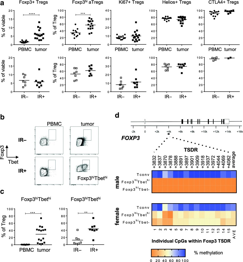
Fig. 3.
Tumor infiltrating Tbet– and Tbethi CD4+CD25+CD127-Foxp3hi cells are bona fide activated Tregs. Freshly dissociated OPSCC tumor tissue was analyzed by 12-parameter flow cytometry analysis with antibodies directed against CD3, CD4, CD8, CD25, CD127, Foxp3, CD45RA, Ki67, Helios, CTLA-4 and Tbet. a Scatter plots displaying the percentage of CD4+CD25+CD127–Foxp3+ Tregs as percentage of viable cells, and the percentage of Foxp3hiCD45RA–, Ki67+, Helios+ and CTLA4+ Tregs of percentage CD4+CD25+CD127–Foxp3+ Treg within PBMC (closed circles) and tumor (closed squares; top panel) samples and within immune response-negative (IR-; open squares) and IR+ (closed squares; bottom panel) tumors of 15 OPSCC patients. b Representative examples of the Foxp3 / Tbet staining within PBMC (left) and tumor (right) of a IR– (top) and IR+ (bottom) OPSCC patient are shown. Cells depicted were first gated for viable and single cells, and further gated for expression of CD3, CD4, CD25, absence of CD127 and expression of Foxp3. c Scatter plots displaying the proportion of Foxp3hiTbethi Tregs as percentage of viable cells (left), and as percentage of CD4+CD25+CD127–Foxp3+ Tregs within OPSCC PBMC (closed circles) and tumor (closed squares; left panel) and within IR– (open squares) and IR+ (closed squares; right panel) OPSCC tumors. * p < 0.05, ** p < 0.01, *** p < 0.001 and, **** p < 0.0001. d Heat map plots displaying the methylation percentages of the individual CpG sites within the Foxp3 Treg cell-specific demethylated region (TSDR) and the average (AVE) of the 15 CpG sites for CD4+CD14–CD25–CD127+Foxp3– conventional cells (Tconv), CD4+CD14–CD25+CD127–Foxp3hiTbethi and Foxp3hiTbet– cells from a male (top) and female (bottom) donor. Populations were isolated from dissociated OPSCC tumors following FACS sorting on a BD FACS Aria II. Percentage methylation is depicted in color-code as depicted in the legend. Treg cells are partially demethylated in the female donor due to Foxp3 methylation on the inactive X-chromosome

