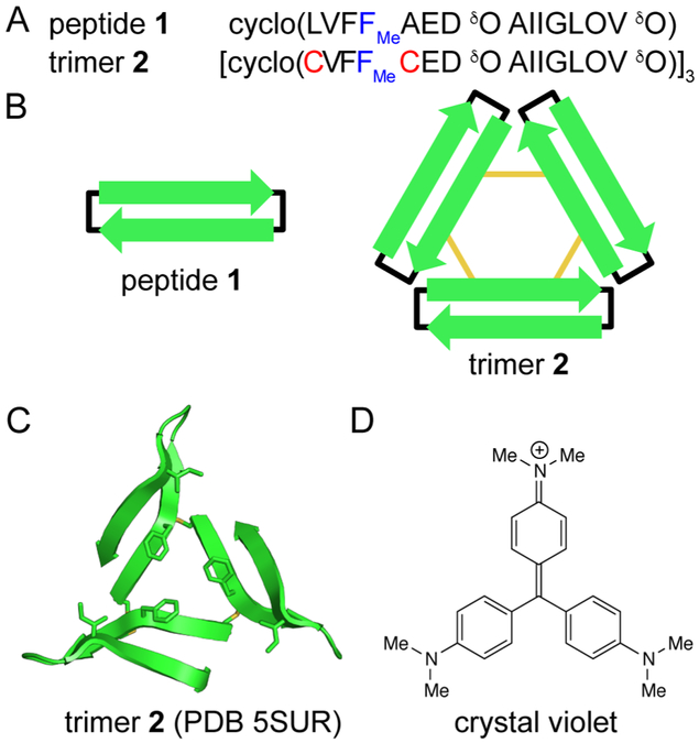
Figure 1:
Chemical models of oligomers formed by Aβ. (A) Amino acid sequences of peptide 1 and trimer 2. (B) Cartoon of peptide 1 and trimer 2. Black lines represent δ-linked ornithine turn units; yellow lines represent disulfide bonds. (C) X-ray crystallographic structure of trimer 2 (PDB 5SUR). The side chains of Cys17, Phe20, Cys21, and Ile31 are shown as sticks. (D) Chemical structure of crystal violet.

