Figure 3:
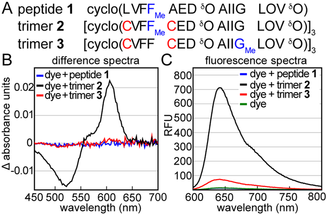
Interaction of other Aβ-derived peptides with crystal violet. (A) Amino acid sequences of peptide 1, trimer 2, and trimer 3. Residues bearing N-methyl groups are highlighted in blue. (B) Difference absorbance spectra of 25 μM crystal violet with 75 μM peptide 1, 25 μM trimer 2, or 25 μM trimer 3. Difference spectra are calculated by subtracting the absorbance spectrum of 25 μM crystal violet from the absorbance spectra of the mixed samples. (C) Fluorescence spectra of 25 μM crystal violet or 25 μM crystal violet with 75 μM peptide 1, 25 μM trimer 2, or 25 μM trimer 3. Fluorescence spectra were acquired with a 550 nm excitation wavelength. Crystal violet exhibits near-baseline fluorescence alone or with peptide 1. Experiments were performed in buffer comprising 50 mM sodium acetate and 50 mM acetic acid (pH 4.6).

