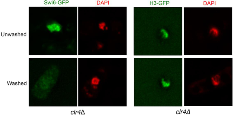
Figure 2. In situ chromatin-binding assay for clr4Δ cells expressing either Swi6-GFP or H3-GFP.
Swi6-GFP is diffused in the nucleus in clr4Δ cells before washing with Triton X‐100 (top panel). The signal can be readily removed upon washing with the detergent (bottom panel). In contrast, the H3-GFP signal in clr4Δ cells is retained in the nucleus before (top panel) or after (bottom panel) Triton X‐100 extraction, indicating that H3-GFP is stably bound with the chromatin. Cells are counterstained with DAPI (red) to visualize the nucleus.

