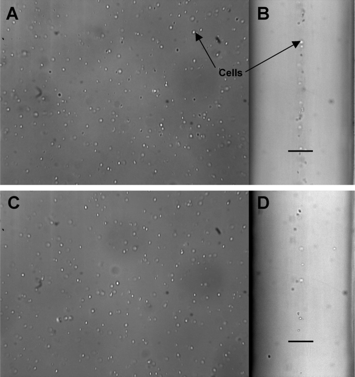
Figure 4.
Brightfield microscope images of cells within the device (10 × magnification). A: Face (y-direction) view near the channel inlet. B: Profile (z-direction) view near the inlet showing the cell stream flowing between two wash streams. Scale bar denotes 115 μm. C: Face view near the outlet. D: Profile view near the outlet.

