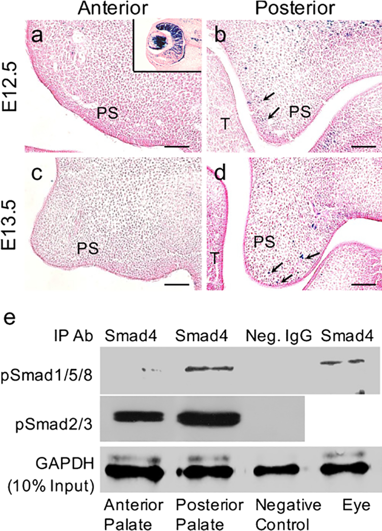
Fig. 2.

Canonical BMP signaling is operating in the posterior but not in anterior portion of the developing palatal shelves. a-d X-gal staining of the anterior (a, c) and posterior (b, d) palate from BRE-Gal reporter mice at embryyonic day 12.5 (E12.5; a, b) and E13.5 (c, d). Black arrows indicate positive X-gal Staining (T tongue, PS palatal shelf). Insert in a Eye tissue as a positive control for X-gal staining. Bars 50 μm. e Western blot showing pSmad1/5/8-Samd4 and pSmad2/3-Samd4 complexes in palatal shelves. Eye tissues were included as positive controls for pSmad1/5/8-Samd4 complexes (IP Ab immunoprecipitation antibody, Neg. IgG negative control IgG, GAPDH D-glyceraldehyde-3-phosphate dehydrogenase)
