Fig. 3.

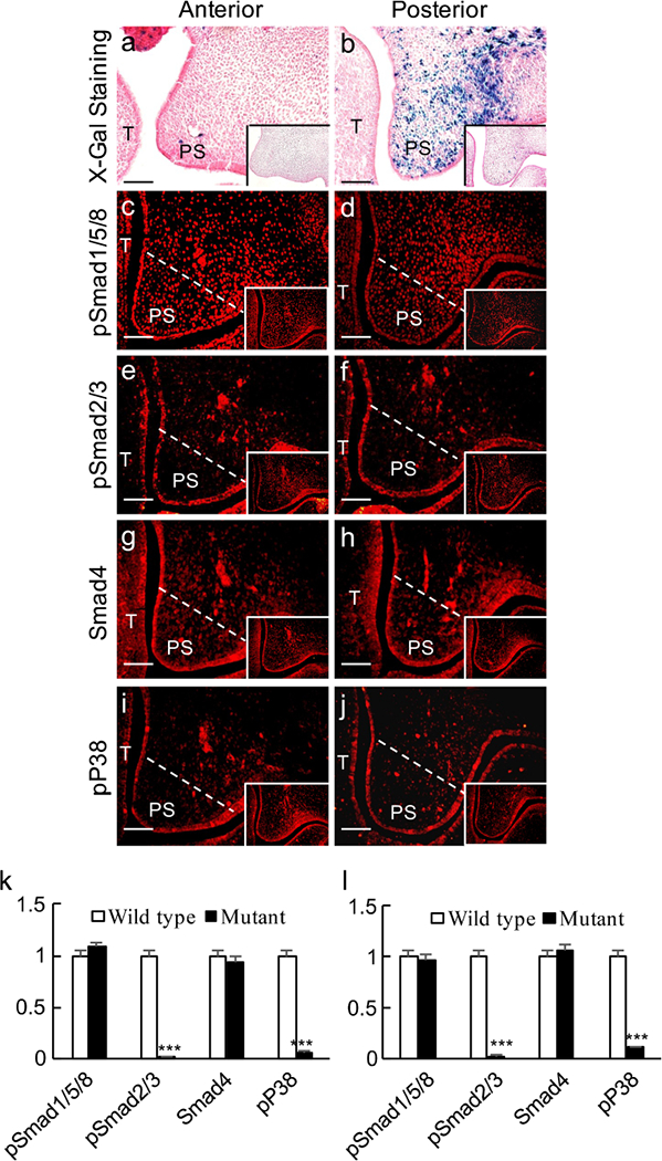
Inactivation of Tgfbr2 in the palatal mesenchyme enhances canonical BMP activity. a, b X-gal staining of the anterior (a) and posterior (b) palate from an E13.5 Wnt1-Cre;Tgfbr2f/f;BRE embryo. c-j Immunofluorescence staining of pSmad1/5/8 (c, d), pSmad2/3 (e, f), Smad4 (g, h) and pP38 (i, j) in the anterior (a, c, e, g, i) and posterior (b, d, f, h, j) palate from E13.5 Wnt1-Cre;Tgfbr2f/f;BRE embryos. White lines demarcate the palatal region for quantifying the immunofluorescence intensity (T tongue, PS palatal shelf). Inserts Wild-type controls. Bars 50 μm. k, l Statistical comparison of immunofluorescence intensity. ***P < 0.001
