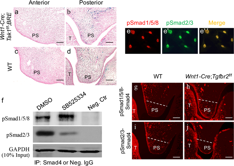
Fig. 4.

Inhibition of TGF-β/Smad2/3 signaling induces pSmad1/5/8-Smad4 complex formation. a-d X-gal staining of the anterior (a, c) and posterior (b, d) palatal shelves from E12.5 Wnt1-Cre;Tak1f/f;BRE (a, b) and wild-type (c, d) embryos (T tongue, PS palatal shelf). e–e” Immunofluorescence showing the co-localization of pSmad1/5/8 and pSmad2/3 in cultured primary posterior palatal mesenchymal cells from E13.5 wild-type embryo. f Western blot showing a significantly increased level of pSmad1/5/8-Smad4 complexes after treatment with SB525334 (Neg. Ctr negative control, IP immunoprecipitation, Neg. IgG negative control IgG). g-j In situ proximity ligation assay shows pSmad1/5/8-Smad4 (g, h) and pSmad2/3-Smad4 (i, j) complexes in the posterior palate of E13.5 wild-type mice (g, i) and Wnt1-Cre;Tgfbr2f/f mice (h, j). Bars 50 μm
